-
Cancer
PET/MRI biomarkers guide personalized treatment for patients with pancreatic cancer
Share this:


Positron emission tomography (PET)/MRI has changed how Mayo Clinic specialists treat patients with pancreatic cancer.
Mayo Clinic pioneered the use of PET/MRI to manage pancreatic cancer. Now a new study by Ananya Panda, M.B.B.S., and colleagues systematically evaluates the strengths of PET/MRI for pancreatic ductal adenocarcinoma (PDAC).
In less than five years, Mayo's PET/MRI practice has grown from a brand-new technology with unknown promise to the most important imaging available to treat pancreatic cancer.
Mayo added its first PET/MRI scanner in 2015 in Rochester, with additional scanners added in Arizona in 2016 and Florida in 2017. Pancreatic cancer imaging has been one of the primary drivers behind significant growth in PET/MRI.
"It has revolutionized what we do," says Mark Truty, M.D., a hepatobiliary and pancreatic surgeon at Mayo Clinic. "The PET/MRI has advantages over a regular PET/CT scan because it combines both a higher-sensitivity PET scanner with a high-resolution MRI, and they do it at the same time. So you could see all the anatomy, along with the tumor and how much of the tumor is alive." Dr. Truty is a co-author on the study.
During a conference presentation, Dr. Truty had an aha moment. "I heard the presenters talking about PET/MRI, and I said: 'Wait a second, that's exactly what I had been looking for in caring for patients with pancreatic cancer. Let's use it. And now the No. 1 indication for PET/MRI at Mayo Clinic is pancreas cancer."
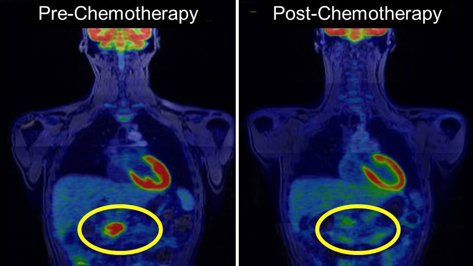
Dr. Truty, a world-renowned surgeon who specializes in pancreatic cancer care, says surgery offers patients the best chance at a longer life. But these operations are more likely to succeed after chemotherapy. PET/MRI helps the team know whether the current chemotherapy regimen is working or if another is needed, and when there is enough dead tumor tissue for a surgery to be effective.
"We can see the tumor die on the PET/MRI scan images," Dr. Truty says. "From the patient perspective, if you're going to get chemotherapy that has side effects, you better be sure it's doing what we think it's doing. And that's what the PET scan allows us to determine much better than a regular CT scan."
PET/MR images then indicate when surgery can be performed to the greatest effect, Dr. Truty says. "We know if the tumor is dead after chemotherapy, then an operation will allow patients to live longer, compared to patients whose tumors are still alive at the time of surgery.
In their American Journal of Roentgenology study, "Borderline Resectable and Locally Advanced Pancreas Cancer: FDG PET/MRI and CT Tumor Metrics for Assessment of Neoadjuvant Therapy Pathologic Response and Prediction of Survival," the authors sought to assess pathologic tumor response to emergent pancreatic cancer therapies and predict overall survival using PET/MRI and CT data.
PET/MRI combines high-quality and simultaneous molecular and anatomic imaging capabilities to address one of the clinical needs in pancreatic cancer care. It provides a noninvasive selection tool to decide whether a highly morbid and expensive surgery will benefit the patient in the long run, and whether a timely switch to alternate treatment regimens could optimize a patient's chances for favorable long-term outcomes, the authors write.
Ajit Goenka, M.D., a Mayo Clinic radiologist and senior author of the study, says the realization of PET/MRI's potential in pancreatic cancer has led to a major shift in how it is used, providing surgeons like Dr. Truty with a clearer picture of whether therapies are working in patients and guiding next steps..
With the metrics obtained through PET/MRI and CT, the researchers found that Mayo Clinic specialists could tailor care to each patient's disease profile.
"We've probably done over 1,000 PET/MRIs. In this particular study, we found that the PET/MRI was much better in predicting how cancer would respond than regular CT scans or blood tests that we normally would use. And that's how this is really changing the practice," Dr. Truty says. "The first thing I want to see after my first visit with the patient after they get their first rounds of chemotherapy is what is the PET/MRI telling me? And then we make significant treatment decisions based on that PET scan."
The study authors are Dr. Panda; Dr. Truty; Dr. Goenka; Timothy Kline, Ph.D.; Matthew P. Johnson; Eric Ehman, M.D.; Garima Suman, M.D.; Bradley Kemp, Ph.D.; Geoffrey Johnson, M.D., Ph.D.; Thorvardur (Thor) Halfdanarson, M.D.; Sudhakar Venkatesh, M.D.; Jeff Fidler, M.D. ― all from Mayo Clinic ― as well as Ishan Garg, M.B.B.S., Maulana Azad Medical College, and Deema Anaam, M.B.B.S., who is pursuing a radiology residency at the University of California San Diego.
###
About Mayo Clinic
Mayo Clinic is a nonprofit organization committed to innovation in clinical practice, education and research, and providing compassion, expertise and answers to everyone who needs healing. Visit the Mayo Clinic News Network for additional Mayo Clinic news.
Media contact:
- Ethan Grove, Mayo Clinic Public Affairs, newsbureau@mayo.edu